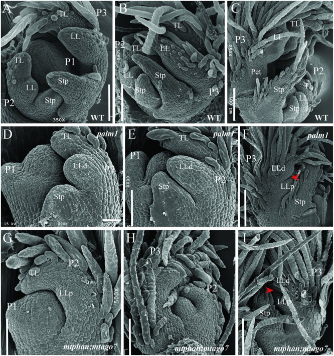
FIGURE 7.

Scanning electron microscopic analyses of leaf primordia development in WT, and palm1 and mtphan;mtago7 mutants. SEM images of leaf development in WT (A–C), palm1 (D–F), and mtphan;mtago7 (G–I). Some trichomes were removed in order to view the boundary between LLd and LLp (I). Arrowheads (F,I) denote the boundary between LLd and LLp. P, plastochron; TL, terminal leaflet; LL, lateral leaflet; Stp, stipule; LLd, distal lateral leaflet; LLp, proximal lateral leaflet.
