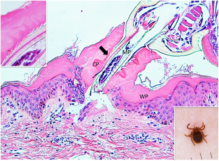
Fig. 1.
(case 1). Histopathology and dermoscopy. Mouthparts (MP) penetrate the epidermis. They are located within conical part (CP) of the external cement which forms a tube. Wing-like part (WP) of it spreads over the epidermis. An arrow indicates serrated structures. Hematoxylin and eosin stain. Original magnification: x100.
Insert in the left upper corner. Magnified serrated structures. Hematoxylin and eosin stain. Original magnification: x200.
Insert in the right lower corner. Dermoscopic feature of an attached tick.

