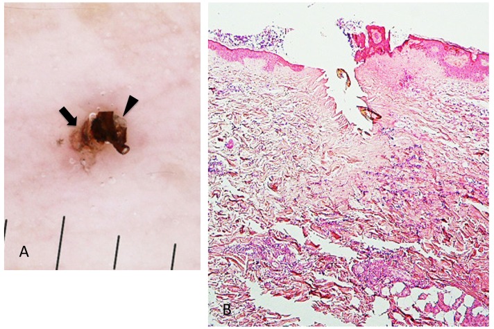
Fig. 3.
(case 2). A (left). Dermoscopic feature. A part of the mouthparts (arrowhead) gets into the external cement (arrow). B (right). Histopathology. There is a V-shaped deep cleavage which is referred to as feeding cavity. There is slight perivascular lymphohistiocytic infiltrate in the entire dermis. Hematoxylin and eosin stain. Original magnification: x20.

