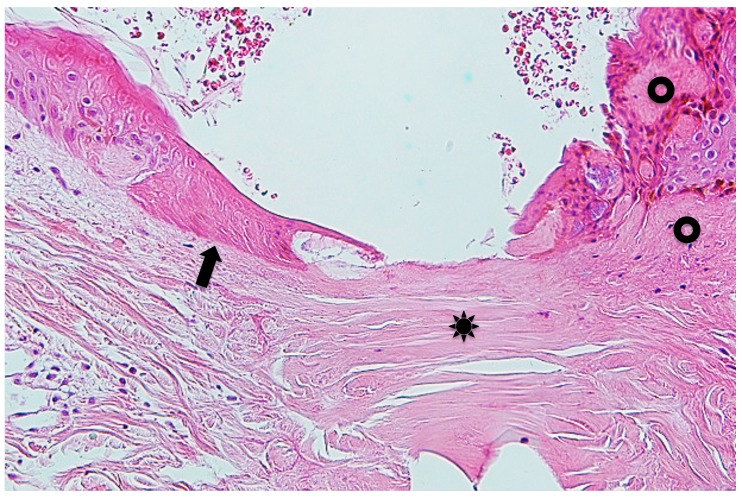
Fig. 6.
Manified Fig. 4B. Arrow indicates massive epidermal cells which undergo coagulative necrosis. Asterisk indicates collagen bundles in the coagulative necrosis area. Two circles indicate papillary and subpapillary areas which show homogeneous, prominent eosinophilic appearance, indicating existence of the permeable toxic agents in the tick saliva. Hematoxylin and eosin stain. Original magnification: x100.

