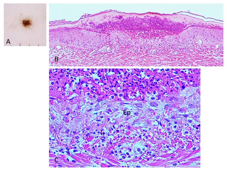
Fig. 9.
(case 3). Dermoscopy and histopathology. A (left upper corner). Dermoscopic feature. There is a brown-black, irregularly shaped square structure. B (top). There is a crust over the epidermis. The crust consists of the wing-like part of the external cement and the underlying aggregated infiltrating inflammatory cells. Hematoxylin and eosin stain. Original magnification: x50. C (bottom). Magnified Fig. 9B. There are numerous erythrocytes and the infiltrating inflammatory cells over the epidermis (Ep). Dermoepidermal junction is indistinct due to the extravasated erythrocytes and the infiltrating inflammatory cells. Hematoxylin and eosin stain. Original magnification: x200.

