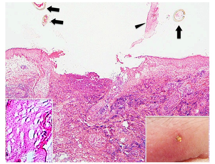
Fig. 17.
(case 6). Histopathology and clinical feature. The epidermis and the underlying dermis are lost, resulting in ulcer formation. Arrows indicate sectioned mouthparts. Arrowhead indicates the epidermis peeled off. Hematoxylin and eosin stain. Original magnification: x20. Insert in the left lower corner. Magnified the epidermis peeled off in Fig. 17. Hematoxylin and eosin stain. Original magnification: x200. Insert in the right lower corner. Clinical feature. There is a part of the tick body which is surrounded by erythema and marked edema on the left upper eyelid.

