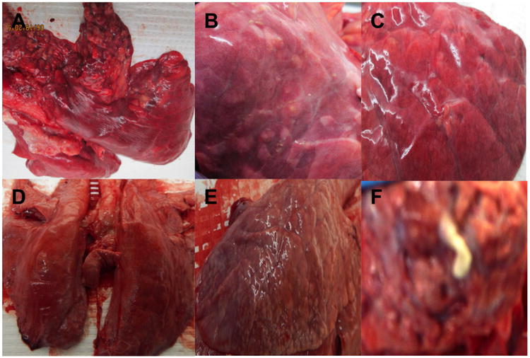
Figure 1. Gross pathology.

Representative gross lesions found in the lungs of pigs from group 1 (A, B, C) and group 2 (D, E, F) demonstrating heterogeneity of TB-compatible lesions. (A) numerous soft and calcified disseminated lesions in apical lobe of pig 59; (B and E) few disseminated soft lesions in one lobe of pig 67; (C-D) very few lesions of pig 76; (F) caseous matter from a lesion collected from pig 94 in group 2 (BSL3 image out of focus but only one available).
