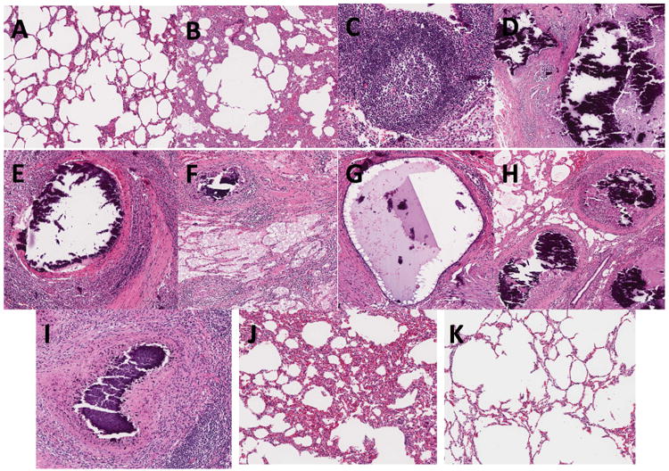
Figure 4. Histopathology.

Representative H&E stained tissue sections of pigs in group 1 (AH) and group 2 (I-K) at 36 and 11 weeks post-challenge of aerosol Mtb strain HN878, respectively. (A-B; J-K) Lung sections from samples obtained in uninvolved areas of the lungs showing low (A,K) or high (B and J) thickening of lung parenchyma; (C) TB compatible granulomas showing sheets of lymphocytes, macrophages, foamy cells and neutrophils; (D-I) TB compatible necrotic granulomas with calcified center, lymphocytes, rim of foamy cells and surrounding fibrosis in the mediastinal lymph node (D, F, H) and lung tissue (E-G and I); (D-H) multifocal necrotic granulomas in lymph nodes.
