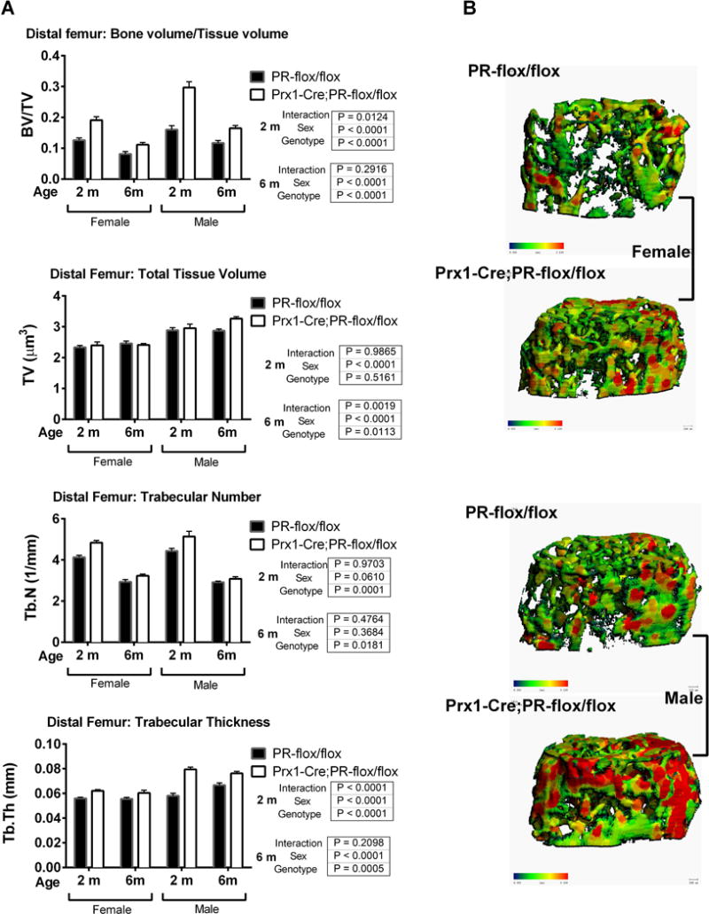
Figure 3. Prx1; PRcKO mice developed high bone mass at the distal femurs in both sexes.

(A). Distal femurs from 2- and 6-month-old female and male Prx1; PRcKO mice were scanned with microCT. Bone volume (BV), total volume (TV), BV/TV, trabecular number (Tb. N), and trabecular thickness (Tb. Th) were compared between wild type and mutant mice. (B). Three-dimensional images were reconstructed for trabeculae at the distal femurs of 2-month-old mice.
