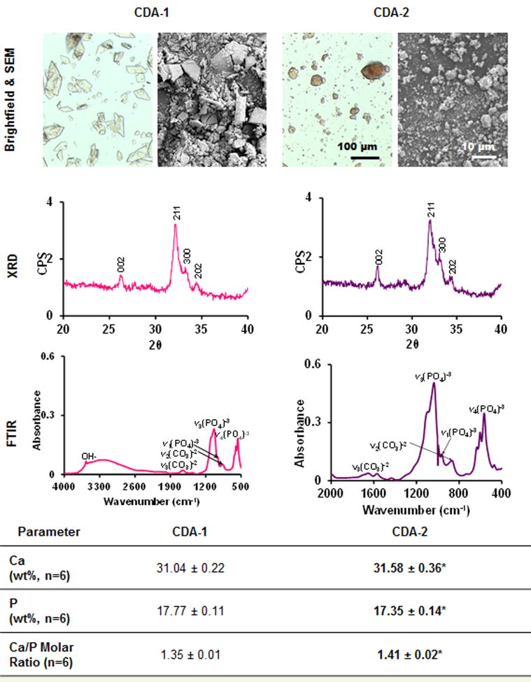
Figure 1. Ceramic characterization.
Brightfield (20×, n=2) and scanning electron microscopy (1000×, n=2) reveal differences in particle shape between calcium phosphate powders. Characteristic phosphate bending curves as well as hydroxyl and carbonate peaks were identified with FTIR (n=2). XRD (n=2) shows that both powders are poorly crystalline. CDA-1 was found to have significantly lower calcium/phosphorus ratio than CDA-2 as determined via inductively coupled plasma analysis (n=6, difference between CDA-1 and CDA-2 *p<0.05).

