Fig. 3.

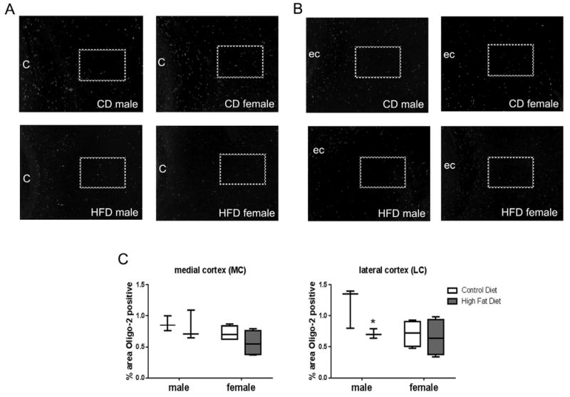
Brain tissue sections from PN 7 pups were stained for oligo-2 protein. Fixed brain sections were incubated with oligo-2 antibody followed by a fluorescent secondary antibody (AlexaFluor 488 nm). Medial cortex (A) and lateral cortex (B) fluorescence were quantified as percent positive staining to total area using threshold settings in Image J software. “C” denotes cingulum; “ec” denotes external capsule; and boxes denote areas quantified. Plot box indicates 25–75 percentile, line indicates median, and whiskers are minimum and maximum values. Data were analyzed by two-way ANOVA, no statistical effects were observed. Multiple t-test indicated a difference between CD and HFD in males in the lateral cortex, “*”statistically different than CD same sex, n = 3–4.
