Fig. 9.
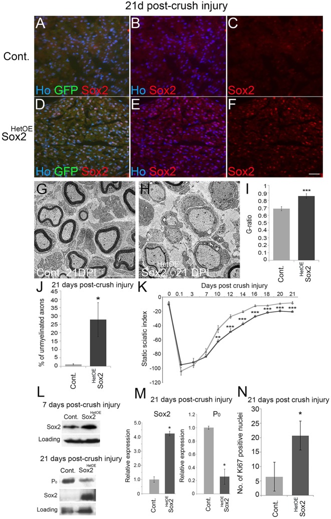
Sustained Sox2 expression results in hypomyelination and reduced functional recovery following nerve injury. (A-F) Immunolabelling of distal nerve sections 21 days post-crush injury (DPI) revealed that nerves from control animals no longer expressed Sox2 at this time point (A-C), whereas Sox2 (and GFP) levels remained elevated in the distal sciatic nerves of injured Sox2HetOE mice (D-F). Scale bar: 40 µm. (G,H) TEM pictures of distal nerve sections at 21 DPI revealed that axons in control nerves are remyelinated (G), whereas few axons are remyelinated in Sox2HetOE nerves (H). Scale bar: 5 µm. (I) G-ratio measurements revealed that Sox2HetOE sciatic nerves were significantly hypomyelinated compared with control nerves at 21 DPI. (J) There is a significant increase in the percentage of unmyelinated axons in Sox2HetOE nerves compared with controls at 21 DPI. Two-sided two-sample Student's t-test; data from n=3 mice of each genotype. (K) Quantification of functional recovery by static sciatic index (SSI). Control, n=5; Sox2HetOE, n=7. Two-sided two-sample Student's t-test. (L) Western blot showing high levels of Sox2 at 7 and 21 DPI and a reduction in P0 expression in Sox2HetOE mice at 21 DPI. (M) Quantification of western blots from L. Two-sided two-sample Student's t-test; data from n=3 independent blots. (N) There is significant increase in the number of Ki67-positive nuclei in distal Sox2HetOE sciatic nerves at 21 DPI. Two-sided two-sample Student's t-test; data from n=3 mice of each genotype. *P<0.05, ***P<0.005.

