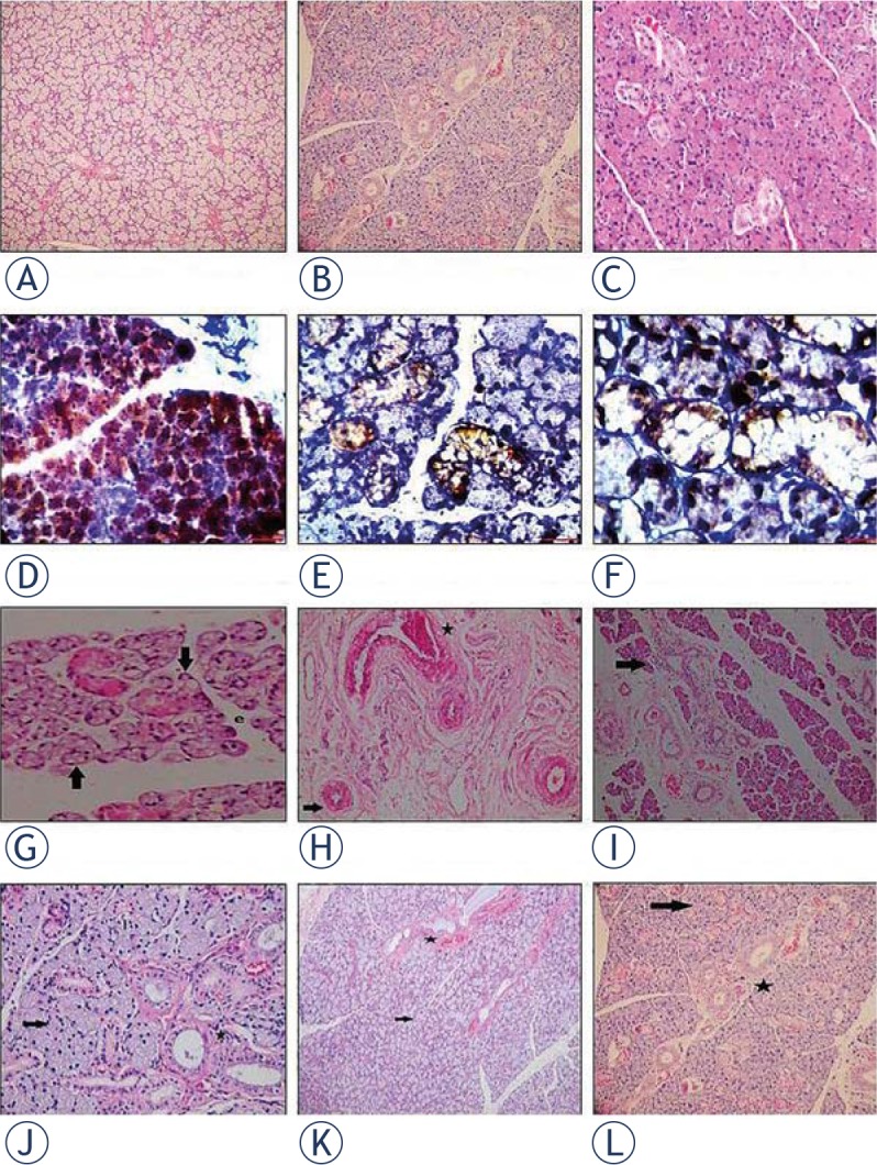
Figure 1.

Histological photographs of salivary gland tissue samples. Pictures on the upper horizontal line show control groups at 1st month evaluation (A; haematoxylin and eosin (H&E)×40), and at 6th month evaluation in different magnifications (B; H&E×200, C; H&E×400). Pictures on the second horizontal line (D to F; ×200 Leica DM 1000) shows the localization of BrdU(+) Adipose Derived Mesenchimal Stem Cells (ADMSC) in the salivary gland (brown stained cells) at 1st month after its intraperitoneal injection in different magnifications. Radioiodine-induced damage in Group 1 is seen in pictures on the third horizontal line (G to I; H&E×400, H&E×400 and H&E×100). Massive vacuolization, necrosis (black arrow) and edema were shown in G minimal vacuolization (black arrow) and periductal fibrosis (black star) in H and lenfosit infiltration (black arrow) in I. Pictures on the lower horizontal line (J to L H&E×400, H&E×200 and H&E×40) indicates the improvements in the histological findings (including vacuolization, fibrosis and edema: black arrow; nearly normal mucous acini, black star; nearly normal ductal system) at 6th month evaluation after the late ADMSC administration in Group 6.
