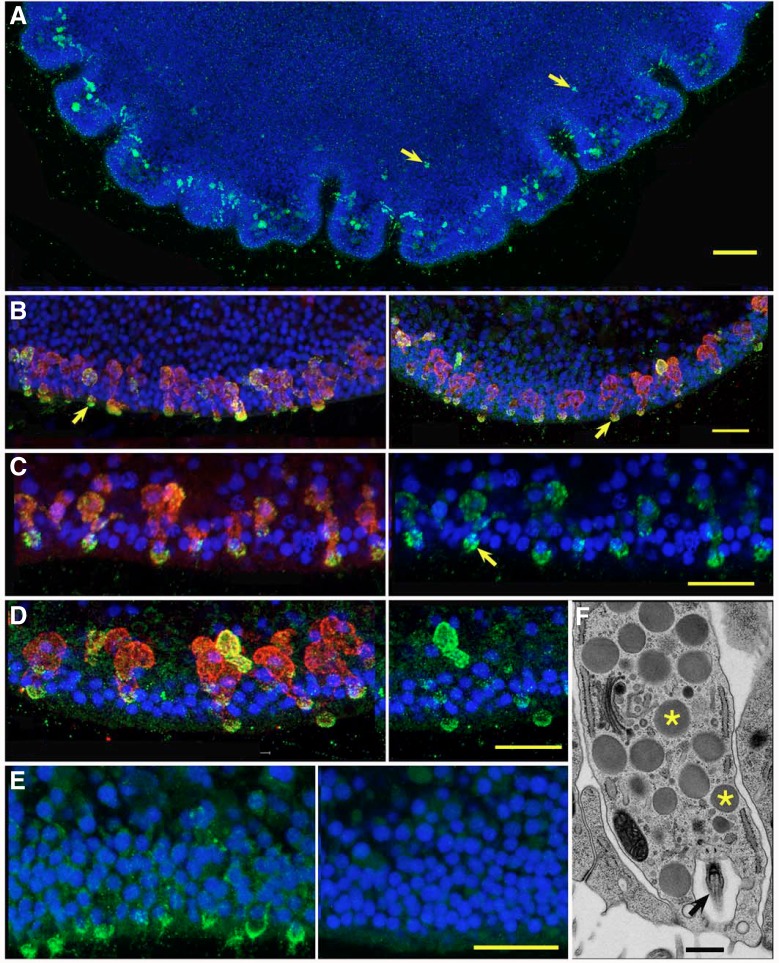
Fig. 4.
ELP and TaELP propeptide are expressed in secretory cells. Confocal (A–E) and transmission electron microscopy (TEM) (F) images of secretory cells. (A) Enface view of a Trichoplax labeled with anti-endomorphin-2. Labeled cells are prevalent around the edge but also present, although dimmer, further toward the center (arrows). (B) Double labeling for endomorphin-2 (left panel; green) or TaELP propeptide (right panel; green) and complexin (red) in secretory cells near the edge of the animal. Complexin staining extends throughout the cells while endomorphin-2 and TaELP propeptide staining is concentrated in cell endings at the exterior surface (arrows). (C,D) Enlarged views of cells double-labeled for endomorphin-2 (C, left panel) or TaELP propeptide (D, left panel) and complexin. Corresponding green channel images (right panels) reveal that label for endomorphin-2 is packaged in larger granules (arrow) than the puncta labeled by the propeptide antibody. (E) Comparison of staining for TaELP propeptide and matched preimmune control showing lack of TaELP staining in control. (F) Serial thin section from freeze-substituted animal showing a secretory cell packed with secretory granules (asterisks) and bearing a cilium (arrow) in a deep pocket at the exterior surface. B–D were captured with an AiryScan detector. Scale bars: A, 50 µm; B–E, 10 µm; F, 0.5 µm.

