
Figure 7.
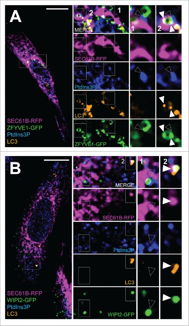
In situ tracking of pre-autophagosome structures at the ER membrane. HeLa cells were transiently transfected with RFP-SEC61B and with GFP-ZFYVE1 or GFP-WIPI2 and were starved in EBSS medium for 15 min. (A) Confocal microscopy analysis of RFP-SEC61B (magenta), LC3 (orange), PtdIns3P (detected by GST-2xFYVE fluorescence, artificially shown in blue) and GFP-ZFYVE1 or (B) GFP-WIPI2 (green). Empty arrowheads indicate SEC61B, PtdIns3P, and ZFYVE1/DFCP1 (or WIPI2) codistribution (early-autophagic PtdIns3P pool). White arrowheads mark SEC61B, PtdIns3P, ZFYVE1 (or WIPI2) and LC3 codistribution (total autophagic PtdIns3P pool). Scale bars: 10 μm.