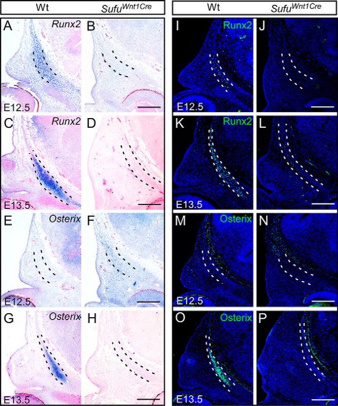
Figure 4.

Expression of the osteogenic markers in development of the frontal bone primordium with in situ hybridization and immunofluorescence analyses. A–H, in situ hybridization showing the expression of Runx2 and Osterix during the frontal bone development in Sufufx/fx;Wnt1-Cre (SufuWnt1Cre) mutants and littermate wild-type embryos (n = 3). I–P, immunofluorescence analysis showing the reduction of Runx2 and Osterix production between E12.5 and E13.5 in Sufufx/fx;Wnt1-Cre (SufuWnt1Cre) embryos compared with littermate wild-type mice (n = 3). Dashed lines outline the presumptive frontal primordium. Scale bars, 200 μm.
