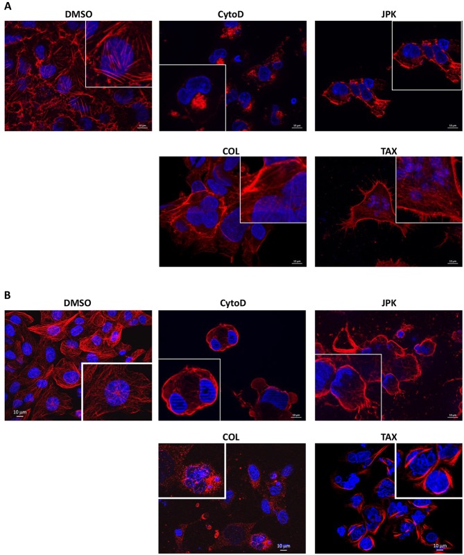
Fig. 1.
Cellular distribution of the cytoskeleton under cytoskeleton inhibitor treatment. 6CFSMEo- cells were incubated with DMSO, cytochalasin D (CytoD), JPK, colchicine (COL) or Taxotere (TAX). After 48 h, the cells were fixed and permeabilized, and incubated with phalloidin to stain actin (A) or with anti-tubulin antibody followed by an Alexa Fluor 594-conjugated secondary antibody (red) for tubulin staining (B). Finally, their nuclei were visualized in blue with Hoechst 33342 stain. These results are representative of two independent experiments.

