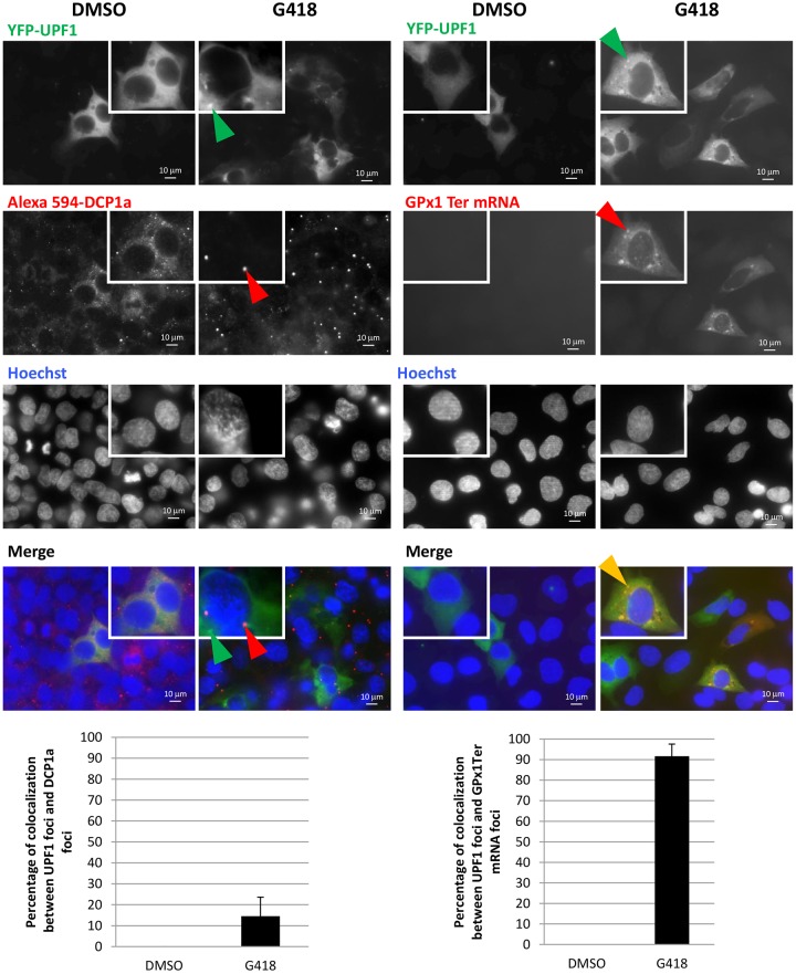
Fig. 6.
G418 causes UPF1 to localize to cytoplasmic foci containing NMD substrates but not the P-body marker DCP1a. 6CFSMEo- cells were transfected with constructs expressing YFP-UPF1 or GPx1 Ter before treatment with G418 at 400 µg/ml for 48 h. Left panel, cells were incubated sequentially with anti-DCP1a antibody and Alexa Fluor 594-conjugated secondary antibody (red). Right panel, cells were incubated with a GPx1 fluorescence in situ hybridization (FISH) probe. The percentage of colocalization between UPF1 and DCP1a (left) or between UPF1 and GPx1 Ter mRNA (right) is presented in histograms at the bottom of the figure. Cells (mean±s.d.; n=10) from three different experiments were counted for each condition. Nuclei were stained with Hoechst 33342 solution (blue). Green arrowheads indicate UPF1 cytoplasmic foci; red arrowheads indicate P-bodies or foci containing GPx1 mRNA; orange arrowheads indicate colocalization foci.

