
Fig. 7.
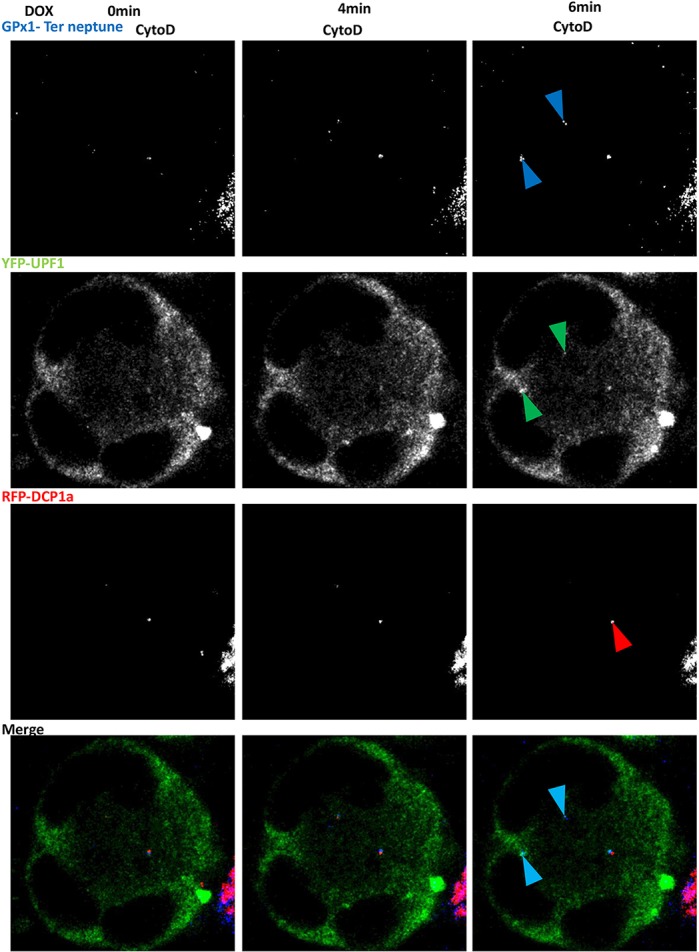
Readthrough GPx1 Ter–Neptune proteins colocalize with UPF1 under cytochalasin D treatment. 6CFSMEo- cells transfected with plasmids encoding YFP–UPF1 (green), RFP–DCP1a (red), and GPx1 Ter–Neptune (blue) were treated with cytochalasin D (CytoD). After 48 h, doxycycline was added to the medium and pictures were taken every 2 min for 1 h under confocal laser scanning microscopy. Pictures taken at 0, 4 and 6 min after adding doxycycline are shown here. Blue arrowheads indicate the GPx1 protein; green arrowheads indicate the UPF1 protein; red arrowheads indicate P-bodies; cyan arrowheads indicate colocalization foci.