Fig. 1.
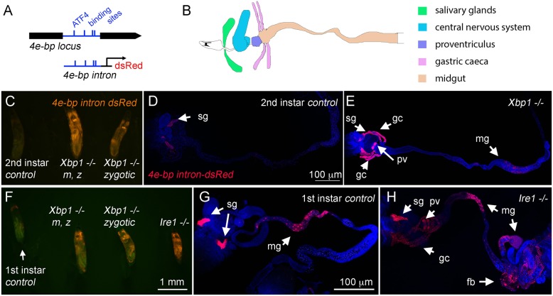
The ATF4-responsive 4E-BPintron dsRed reporter is induced in Xbp1 and Ire1 mutant larvae. (A) A schematic diagram of the 4E-BP locus. The intron is depicted in blue, and the predicted ATF4-binding sites are indicated by vertical lines. The dsRed reporter expression is driven by this intron element (Kang et al., 2017). (B) A schematic diagram of the salivary glands (green), central nervous system (blue), proventriculus (purple), gastric caeca (pink) and midgut (yellow) in the second-instar Drosophila larva. Compare this diagram with images in D,E,G and H. (C–H) 4E-BPintron dsRed expression (red) in the indicated genetic backgrounds. TO-PRO-3 (blue) was used to show the outline of tissues. (C) An intact control of wild-type Xbp1 (left), an Xbp1 maternal zygotic (m, z) mutant (center) and an Xbp1 zygotic mutant (right) at the second-instar stage juxtaposed to each other without dissection. (D) Dissected wild-type second-instar larva with 4E-BPintron dsRed expression primarily restricted to the salivary glands. (E) In the Xbp1ex79−/− background, 4E-BPintron dsRed is strongly induced in tissues beyond the salivary glands, which include the gastric caeca and the proventriculus (arrows). (F) Intact wild-type (left), Xbp1 maternal zygotic (m, z) mutant (left center), Xbp1 zygotic mutant (right center) and Ire1f02170−/− (right) first-instar larvae are juxtaposed. (G) Dissected wild-type first-instar larva showing 4E-BPintron dsRed expression most prominently in the salivary glands. (H) In the Ire1−/− larva, the reporter is also strongly expressed in the proventriculus, gastric caeca, midgut and fat body (indicated with arrows). The scale bar in D applies to D and E; the scale bar in F applies to panels C and F, and the scale bar in G applies to images in G and H. sg, salivary glands; gc, gastric caeca; pv, proventriculus; mg, midgut; fb, fat body.

