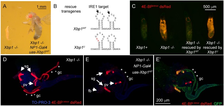
Fig. 2.
Rescue of Xbp1 mutant-associated lethality through the expression of Xbp1 transgene. (A) Xbp1−/− larvae survive only up to the second-instar larval stage (left), whereas Xbp1 mutants expressing transgenic Xbp1 with the NP1-Gal4 driver survive to adulthood (right). (B) The IRE1 target cleavage sequence within the XBP1 mRNA. Arrows point to the sites that are spliced. The wild-type sequence (upper image) and the mutated sequence within the rescue transgene (lower image, in red) are shown. (C–E) The effects of Xbp1 transgene expression driven by NP1-Gal4 on the 4E-BPintron dsRed reporter levels in Xbp1−/− second-instar larvae (red). (C) Non-dissected second-instar larvae of the indicated genotypes. (D,E) 4E-BPintron dsRed signal (red) from dissected second-instar larval tissues. TO-PRO-3 (blue) was used to show the outline of tissues. The strong expression of 4E-BPintron dsRed signal in the Xbp1−/− background (D) is suppressed by the wild-type Xbp1 transgene expression through the NP1-Gal4 driver (E). UAS-GFP was co-expressed with the Xbp1 transgene, and GFP signal (green) marks the NP-Gal4 active tissues, which includes the gastric caeca (asterisks) and the midgut (E′), and 4E-BPintron dsRed signal is specifically suppressed in those domains. The scale bar in E′ applies to D and E. sg, salivary glands; gc, gastric caeca; pv, proventriculus; fb, fat body.

