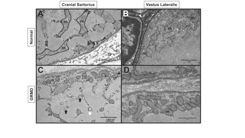
Figure 7.
GRMD dog muscle had abnormal mitochondria (m). Transmission electron microscopy (TEM) of normal and GRMD cranial sartorius (CS) and vastus lateralis (VL) at 6 to 12 months of age. A: Normal CS with intermyofibrillar mitochondria. B: Normal VL with numerous subsarcolemmal mitochondria C: GRMD CS with reduced size and density of subsarcolemmal and intermyofibrillar mitochondria, swollen (white arrow) mitochondria and dilated sarcoplasmic reticulum (black arrows). D: GRMD VL with reduced subsarcolemmal mitochondria.

