
Figure 1.
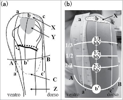
Anatomical structure of the left shoulder (a) and locations of 4 examined sites in a living body (b). A: The upper end of the anterior axillary line, B: the upper end of the posterior axillary line, a: the anterior edge of the mid-acromion lateral border, C: Deltoid tuberosity for attachment of the deltoid muscle, b: the midportion of the mid-acromion lateral border, c: the posterior edge of the mid-acromion lateral border, a’: the intersection between the perpendicular line drawn from the anterior edge of the mid-acromion lateral border and line AB, b’: the intersection between a perpendicular line drawn from the mid-acromion lateral border and line AB, c’: the intersection between the perpendicular line drawn from the posterior edge of the mid-acromion lateral border and line AB, X: acromion, Y: subdeltoid/subacromial brusa, Z: humerus, dotted circle: the deltoid muscle, dotted line: the posterior circumflex humeral artery (PCHA), black line below the PCHA: the axillary nerve. (b) One third, half, and two thirds of bb’ are marked on the skin. 1/3 bb’, 1/2 bb’, 2/3 bb’, and b’ are the sites examined in the present study.