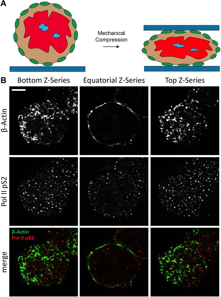
Fig. 7.
Utilization of a mechanical microcompressor to visualize proteins of the nuclear lamina. (A) Schematic of mechanical microcompression. (B) From left to right, deconvolved images of the bottom, equatorial and top confocal sections of a representative nucleus stained for β-actin (green) and Pol II pS2 (red). Confocal z-series of representative nuclei are shown. Scale bar: 5 μm.

