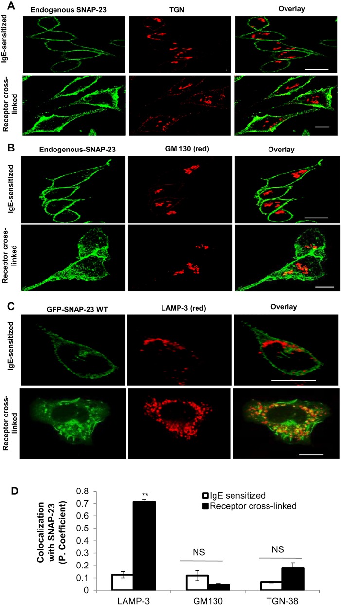
Fig. 2.
SNAP-23 shows significant colocalization with lysosomal granule marker LAMP-3 but not with TGN38 and GM130 in allergen-stimulated RBL MCs. (A,B) The representative confocal images are showing that endogenous SNAP-23 (green, SNAP-23 Ab) does not relocate to the TGN or Golgi membrane [(red, TGN-38 Ab; n=71, IgE sensitized; n=100, receptor cross-linked) and GM-130 Ab (n=100, IgE sensitized; n=70, receptor cross-linked)] after receptor cross-linking of MCs. (C) Representative confocal image is showing the GFP-SNAP-23WT (green) co localization with lysosomal granules (red, granule marker LAMP-3-specific Ab). A high amount of colocalization (yellow, overlay) of GFP-SNAP-23WT with LAMP-3 containing granules was observed after receptor crosslinking of MCs (n=15). In all resting states SNAP-23 resides on plasma membrane with no colocalization with any internal organelle marker. Scale bar: 10 μm. (D) The graph representing the co-localization of SNAP-23 (both endogenous and transfected) with the counterstained markers (above) in terms of Pearson coefficient. Each data point is mean±s.e.m. of three independent experiments (**P≤0.005, Student's t-test, one-tailed distribution).

