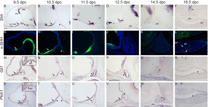
Fig. 1.
Expression of Shh/SHH, Gli1 and Ptch1 during normal pituitary development. In situ hybridisation and immunofluorescence on wild-type mid-sagittal (A-E,G-K,M-Q,S-W) and frontal sections (F,L,R,X) during pituitary organogenesis between 9.5 and 18.5 dpc. (A-F) Shh transcripts are detected in the anterior hypothalamus (AH, arrows), posterior hypothalamus (PH, asterisks) and pharyngeal endoderm (PE, arrowhead) abutting the developing Rathke's pouch (RP). Note the expression in the subventricular zone around the 3rd ventricle (3v). (G-L) SHH immunostaining is observed in a similar pattern. Note the weak SHH signal in the anterior region of RP at 10.5 dpc (H, asterisk). Single SHH+ cells are detected at 18.5 dpc in the AP (arrows) and overlying hypothalamus (asterisk in L). (M-R) In situ hybridisation for Gli1. Note the expression of Gli1 in the developing RP (M-O, arrowheads), periluminal epithelium (P,Q, arrowheads) and marginal zone of the anterior pituitary (R, arrowhead). Within the hypothalamus, Gli1 transcripts are initially detected in the AH (M, arrow), but become restricted to the PH from 10.5 dpc (N-P, asterisks) and subventricular zone of the 3rd ventricle at 18.5 dpc (asterisk in R). (S-X) Ptch1 transcripts are observed in the AH (arrows), PH (asterisks) and around the 3rd ventricle (asterisk in X). Within RP, Ptch1 is initially expressed in the rostral region of the evaginating RP at 9.5 dpc (arrowhead in the inset in S), at the boundary of RP with the oral ectoderm and pharyngeal endoderm (arrowheads in T), and in the ventral region of the pinched-off pouch (arrowhead in U). Subsequently, Ptch1 transcripts are localised within the periluminal epithelium and marginal zone (arrowheads in V-X). AP, anterior pituitary; PP, posterior pituitary; MB, midbrain; HB, hindbrain; IF, infundibulum; OE, oral endoderm; PE, pharyngeal endoderm; VT, ventral telencephalon. Scale bar: 100 μm.

