Figure 3.

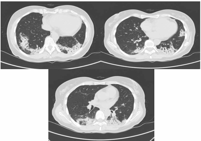
Contrast-enhanced CT scan showing areas of pulmonary consolidation with bilateral distribution and ground-glass opacities.

Contrast-enhanced CT scan showing areas of pulmonary consolidation with bilateral distribution and ground-glass opacities.