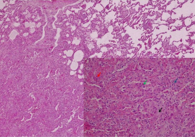
Figure 4.

Surgical lung biopsy (H&E, original magnification 40×). In the lower right corner, in more detail, the characteristic changes of AFOP: alveolar spaces filled by fibrin (red arrow) and macrophages (blue arrow), hyperplasia of type II pneumocytes (green arrow) and foci of fibroblastic proliferation (black arrow) (H&E, original magnification 100×). AFOP, acute fibrinous and organising pneumonia.
