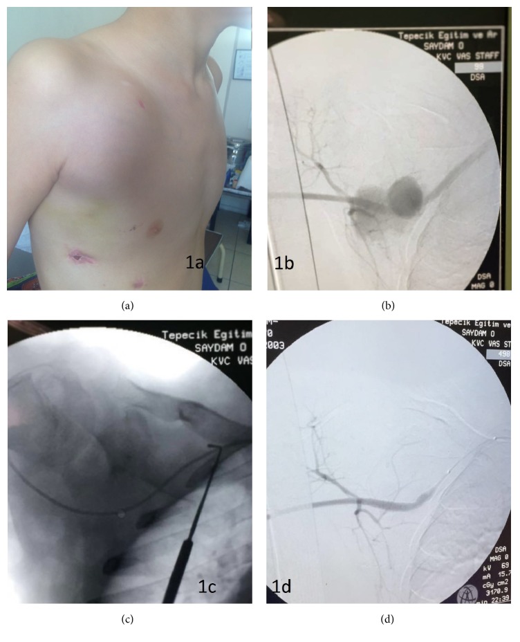
Figure 1.
(a) Large subclavian pseudoaneurysm pulsatile sac with palpable trill. (b) Angiogram of pseudoaneurysm sac originated from the right subclavian artery. (c) Passing the lesion with 0.035 hydrophilic guide wire with the help of 5F straight selective multipurpose diagnostic catheter. (d) Final selective angiogram of the right subclavian artery with a complete hemostasis and preserved subclavian artery branches.

