Fig. 3.
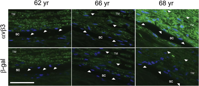
Variable localization intensity of αvβ3 integrin in trabecular meshwork and Schlemm’s canal from 3 different donors. Anterior segment wedges from 3 donors were fixed in 4% p-formaldehyde prior to paraffin embedding. Sections were cut and then labeled with mAbs against either αvβ3 integrin (top) or β-galactosidase (bottom) as a negative control. Arrowheads indicate positive staining that is present in the αvβ3 integrin-labeled sections but absent in the β-galactosidase-labeled sections. Note the variation in labeling intensity observed in tissue from the different donors. Weak αvβ3 integrin labeling is present only along the inner wall of Schlemm’s Canal (SC) within the 62 year old donor tissue. In contrast, αvβ3 labeling is present within SC and along the trabecular meshwork (TM) beams in the other two donors’ tissue sections. However, the labeling is significantly stronger in the 68 year old donor than in the 66 year old donor. Specificity of the anti- αvβ3 integrin antibody (clone [BV3], Abcam, Cambridge, MA) was verified by FACs analysis (not shown) using TM-1 cell lines with known levels of αvβ3 integrin (Gagen et al., 2013). Scale bar = 50 µm.

