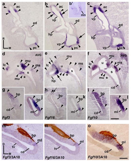
Figure 3. Fgf3 and Fgf16 expression patterns at HH27.
a–l, Horizontal sections through the inner ear at stage HH27, indicated in Figs 5e and 5f. The probes used are indicated in each column. The Fgf10 expression pattern defines the sensory patches (between arrowheads). The Fgf3 transcripts were detected in borders of all cristae (ac, a; pc, a; lc, d). The entire macula utriculi and a part of the macula sacculi showed Fgf3 expression (mu and ms in d). In the cochlear duct, the developing basilar papilla and the macula lagena showed Fgf3 transcripts (bp and ml in g, j). Regarding the Fgf16 gene, strong Fgf16 expression was observed in the areas bordering all Fgf10-positive cristae (long arrows in b, e). The short arrow in e points to the border between the lateral semicircular system and the utricule. In the utricule (u) and saccule (s), parts of their walls were labeled by the Fgf16 expression (u and asterisk in e). The macula neglecta and macula lagena were Fgf16 positive (mn in b′; ml in k). The utricular and saccular maculae were Fgf16 negative (mu and ms in e). The basilar papilla showed some Fgf16-staining cells (short arrow in h). m–o, transverse sections treated with 3A10 immunoreactions, showing the Fgf3/Fgf16-positive macula lagena and the portion of the basilar papilla. For the abbreviations, see the list. Orientation: M, medial; R, rostral. Scale bar = 27 μm in l (applies to a–l) and 20 μm in o (applies to m–o).

