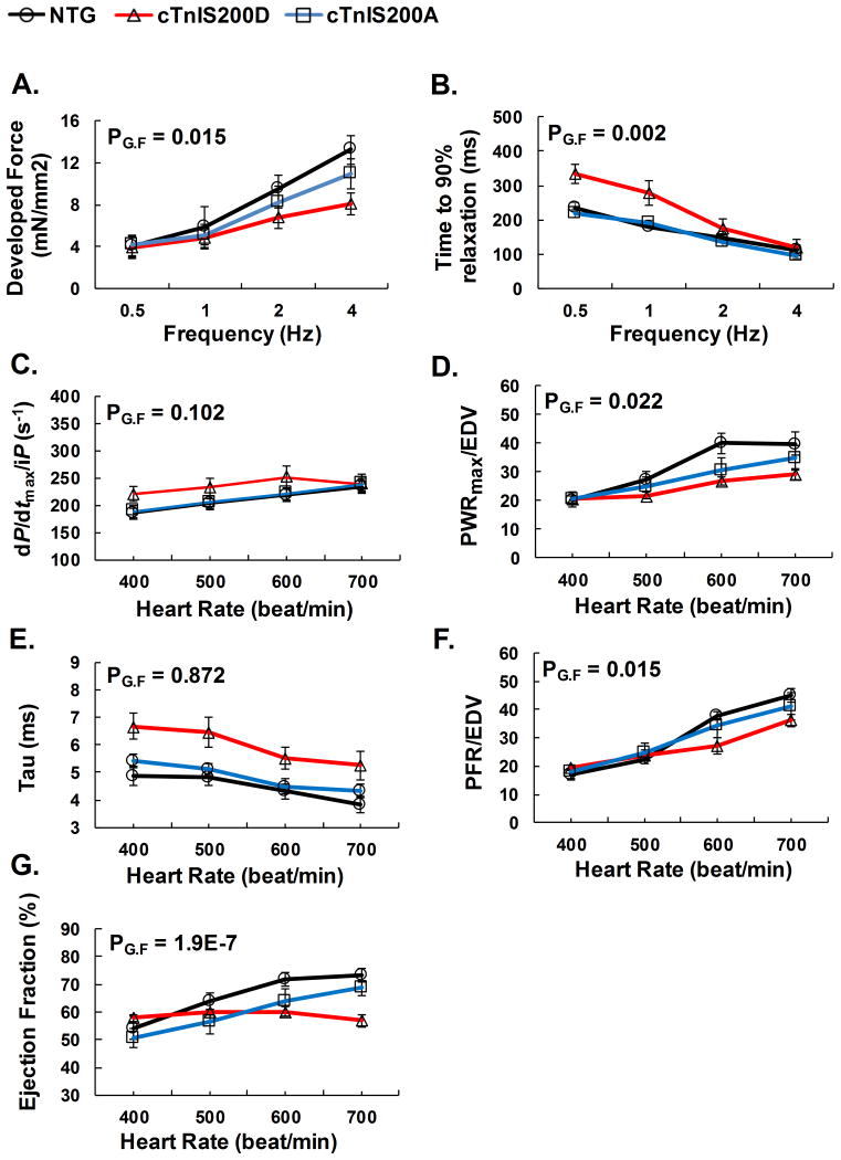
Figure 3.
Cardiac responses to increased pacing in vitro and in situ. A-B. In vitro isometric force measurements on isolated intact trabecular muscle over stimulation frequency 0.5-2 Hz, 1mM external Ca2+, n=5. C-G. In situ hemodynamic study of cardiac function over paced heart rates from 400-700 beats/min. n=5. Explanation of abbreviations is the same as Table 2. PG.F<0.05 indicates significance among the interactions between genotype and pacing frequency. cTnIS200D mice with hyper-phosphorylation mutation depressed positive force-frequency relationship both in vitro and in vivo.

