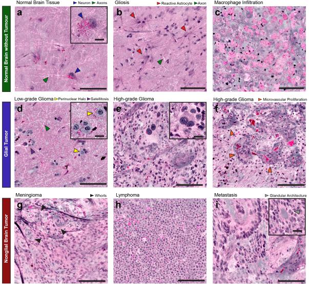
Fig. 3. Imaging of key diagnostic histoarchitectural features with SRH.
(A) Normal cortex reveals scattered pyramidal neurons (blue arrowheads) with angulated boundaries and lipofuscin granules, which appear red. White linear structures are axons (green arrowheads). (B) Gliotic tissue contains reactive astrocytes with radially directed fine protein-rich processes (red arrowheads) and axons (green arrowheads). (C) A macrophage infiltrate near the edge of a glioblastoma reveals round, swollen cells with lipid-rich phagosomes. (D) SRH reveals scattered “fried-egg” tumor cells with round nuclei, ample cytoplasm, perinuclear halos (yellow arrowheads), and neuronal satellitosis (purple arrowhead) in a diffuse 1p19q-co-deleted low-grade oligodendroglioma. Axons (green arrowhead) are apparent in this tumor-infiltrated cortex as well. (E) SRH demonstrates hypercellularity, anaplasia, and cellular and nuclear pleomorphism in a glioblastoma. A large binucleated tumor cell is shown (inset) in contrast to smaller adjacent tumor cells. (F) SRH of another glioblastoma reveals microvascular proliferation (orange arrowheads) with protein-rich basement membranes of angiogenic vasculature appearing purple. SRH reveals (G) the whorled architecture of meningioma (black arrowheads), (H) monomorphic cells of lymphoma with high nuclear:cytoplasmic ratio, and (I) the glandular architecture (inset; gray arrowhead) of a metastatic colorectal adenocarcinoma. Large image scale bars = 100μm; inset image scale bars =20μm.

