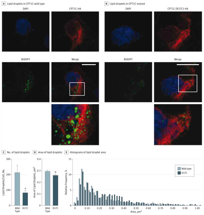
Figure 4. p.Arg37Cys CPT1C Is Associated With Reduced Lipid Droplet (LD) Number in COS7 Cells.
A and B, COS7 cells were transfected with wild-type or mutant hemagglutinin (HA)-tagged CPT1C and stained with HA antibody (clone 16B12, 1:5000; Covance) and BODIPY 493/503 for LDs (0.1 μg/mL; Invitrogen). The bottom panel shows an enlarged view of the boxed area. Scale bar = 20 μm. C and D, Numbers and areas of LDs were counted blindly in an automated fashion and results were derived from a total of 2949 LDs. Results are given as mean (SEM). E, Relative distribution of LD areas in wild-type and mutant CPT1C. Counting of LDs was performed in a blinded and automated fashion using the software Volocity 3D Image Analysis Software (PerkinElmer). The data were analyzed using GraphPad Prism software version 6 (http://www.graphpad.com) and P ≤ .05 was considered significant. DAPI indicates 4′,6-diamidino-2-phenylinodole.
aP < .01.
bP < .05.

