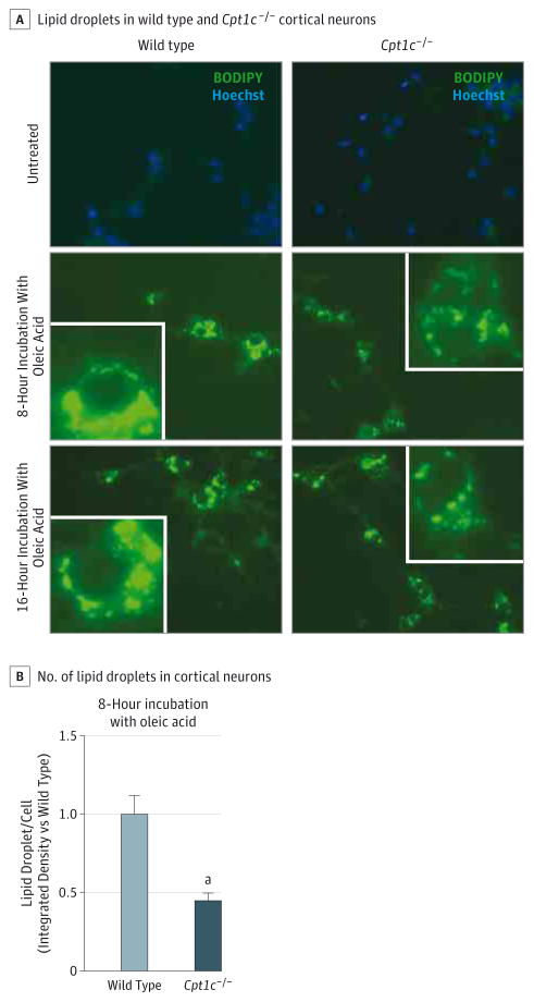
Figure 5. Lipid Droplet Number Is Decreased in Cpt1c−/− Cortical Neurons.
A, Cells were obtained from the cortex of wild-type or Cpt1c−/− E16 embryos and seeded in 48-well plates during 7 days in vitro. For lipid loading, 300μM oleic acid was added for 8 or 16 hours before fixation. Representative images are shown. Images of neural lipids and the nucleus were acquired on a Nikon Eclipse TE-2000E optic microscope using a plan apochromat objective (BODIPY 493/503 and Hoechst dyes [Life Technologies], respectively; original magnification ×20). For quantification, sets of cells were cultured and stained simultaneously and imaged using identical settings. The region of interest was randomly selected using nucleic staining. B, For quantification, the Fiji image processing package was used to determine the integrated density stain per soma in at least 80 cells per condition. The results are shown as the mean of 2 independent experiments ± SEM.
aP < .001.

