
Figure 4.
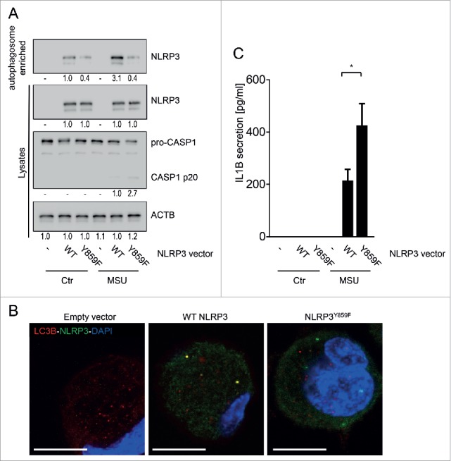
NLRP3 lacking the phosphorylation site is not recruited to phagophores. BMDC from nlrp3−/− mice were transfected with a wild-type (WT) Nlrp3 expression vector, or a Nlrp3 construct where Tyr859 in NLRP3 is replaced with a phenylalanine (Y859F; Y>F) to abolish NLRP3 phosphorylation. The cells were primed for 16 h with upLPS to induce NLRP3 and IL1B expression, before activation with MSU (150 µg/ml) for 6 h. Shown is (A) representative western blot pictures from cell lysates and autophagosome-enriched fractions, (B) confocal microscopy of LC3B (red) and NLRP3 (green) immunostained cells; blue: DNA stained with DAPI, scale bar: 10 μm; and (C) IL1B in the cell culture supernatant. Data are representative for 1 out of 3 independent experiments with 3 replicates each (n = 3), except for confocal microscopy where the experiment has been performed only twice with 3 replicates. Images are representative for at least 5 scanned areas for each depicted condition. Numbers below the western blot pictures show results of densitometric measurements. * = p < 0.05, one-way ANOVA with Bonferroni correction.