
Figure 3.
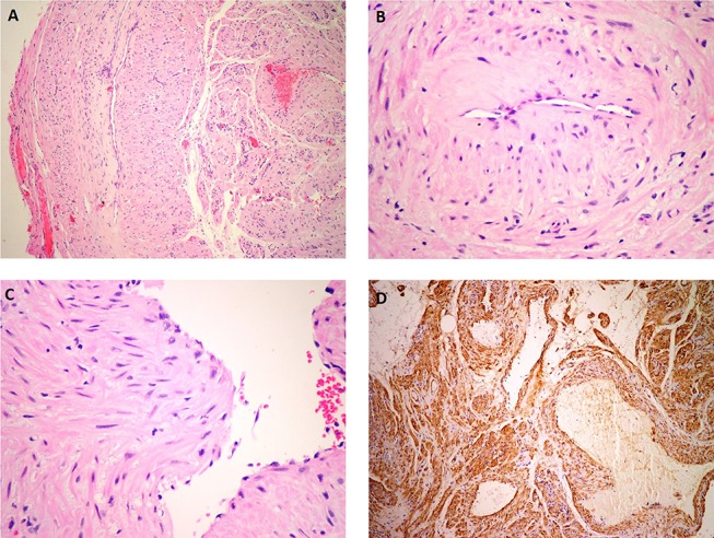
(A) Microscopic examination (H&E) revealed that the mass was well-circumscribed and composed of vascular channels. (B) The vascular channels were surrounded by fascicles of concentrically arranged spindle cells. (C) Tumour cells exhibited eosinophilic cytoplasm and oval nuclei showing no mitotic figures and a minimal degree of pleomorphism. (D) Immunohistochemical examination (SMA) revealed that the spindle cells were strongly positive for smooth muscle actin (SMA). In addition, the walls of vascular spaces were also positive for SMA.