
Figure 9. Swapping the different peptides (red and blue) in and out of the protein binding site.
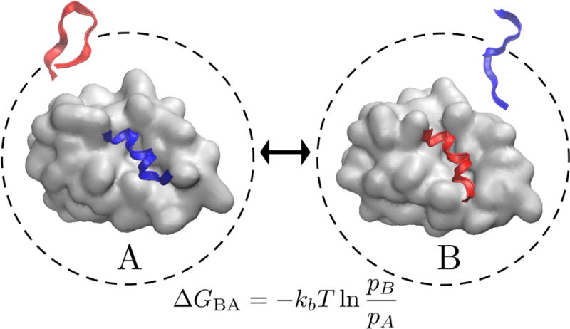
Two possible states (A) in which one peptide (blue structure) is bound to the target protein (gray surface) and the other (red structure) is kept unbound and (B) in which the roles of the peptides are reversed. Each peptide may favor different binding modes. The ratio of the populations between these two cases pB/pA can be related to the relative free energy of binding by the equation given here.