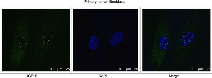
Fig 3. Confocal microscopy analysis of IGF1R nuclear localization in primary human fibroblasts.
Fluorescence confocal microscope imaging of IGF1R in primary human fibroblasts. Cells were seeded in 24-well plates and after 24 hr were fixed and stained for IGF1R with a fluorescent donkey anti-rabbit antibody (green- 488) and DAPI (blue). Results of a representative experiment repeated two times with similar results are shown.

