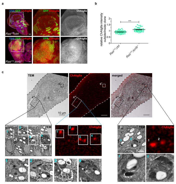
Extended Data Figure 1. NAA is induced in the wing disc and ChAtg8a signal derives from autophagic structures.
a, Representative confocal images of wing imaginal discs of ChAtg8a-animals carrying RasV12 scrib−/− tumours are shown. n = 17 (RasV12ctrl) and n = 19 (RasV12scrib−/−) discs from three independent experiments. b, Quantification of relative ChAtg8a intensities inside and outside clones of indicated genotypes from single confocal sections. Values represent mean and s.e.m. of three independent pooled experiments. n = 24 discs (RasV12ctrl), n = 20 discs (RasV12scrib−/−) ***P < 0.0001 from unpaired two-tailed t-test. c, Transmission electron microscopy, confocal and overlay images correlating fluorescent puncta with organelles at the ultrastructural level (insets are shown enlarged). ChAtg8a-positive areas correlate with autophagosomes and autolysosomes (1–3) and apoptotic cells with autolysosomal profiles (4). Scale bars, 50 μm (a) and 10 μm (c).

