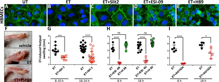
Fig 5. Arf6 and Epac/Rap1 play key roles in mediating EF toxicity in mammalian systems.
(A-E) HBMECs (Human Brain Microvascular Endothelial Cells) stained with an anti pan-Cadherin antibody. (A) The pan-Cadherin (p-Cad) stain clearly delineates cell borders in untreated cells (UT). (B) ET (EF+PA) treatment results in strong reduction in Cadherin staining and loss of AJs. (C) Co-treatment of cells with ET and the Slit2 peptide, which prevents Arf6 activation via induction of ArfGAP activity, fully restores AJs compromised by ET treatment. (D) Co-treatment with the Epac-specific inhibitor ESI-09 partially rescues p-Cad expression at AJs. (E) Co-treatment with the PKA-inhibitor H89 does not appreciably rescue junctional expression of pCad at cell borders but does restore intracellular p-Cad staining (F-I) ET-induced footpad swelling (edema) can be suppressed by pharmacological intervention. (F) ET-injected footpads develop a striking edema. Pre-treatment with ESI-09 consistently blocks this symptom. Pre-treatment with (G) SecinH3, a compound that indirectly suppresses the activation of Arf6 (****p<0.0001, ***p<0.001), and (H) the Epac pathway inhibitor ESI-09, but not the PKA inhibitor H89, abrogate EF-induced edema (****p<0.0001, ns non significant). (I) AG1024, a compound that may indirectly block Rap1 activation, acts as a potent inhibitor of ET-induced edema at early times, but offered more modest protection at a later time point (****p<0.0001, *p<0.05).

