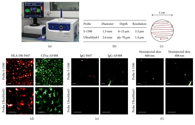
Figure 1.
FCFM imaging of the skin. FCFM system (a). Characteristics of the probes (b). Continuous scanning was performed manually on imaged area that covers a diameter of 1 cm (c). Representative in vivo images of the skin performed 30 min after injection of anti-HLA-DR-F647 or anti-CD1a-AF488 antibodies (d); corresponding isotype controls (e). Untreated skin (f) (n = 3). Scale bars: 50 μm.

