Fig. 6.
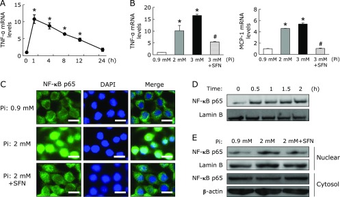
High Pi loading directly increased cytokine/chemokine mRNA expression through the activation of NF-κB in RAW264.7 cells. (A) RAW264.7 cells were treated with Pi (2 mM) for the indicated times (n = 3). (B) RAW264.7 cells were pretreated with SFN (10 µM) for 1 h, followed by treatment with different Pi concentrations for 1 h (n = 3). Relative TNF-α and MCP-1 mRNA levels were determined by quantitative real-time RT-PCR. (C) RAW264.7 cells were pretreated with SFN (10 µM) for 1 h and then exposed to Pi (2 mM) for 30 min. Cells were fixed, permeabilized and stained with NF-κB p65 and DAPI. Magnification bar, 10 µm. (D) RAW264.7 cells were treated with Pi (2 mM) for the indicated times and nuclear protein was extracted. Nuclear NF-κB p65 expression was determined by Western blot analysis. (E) RAW264.7 cells were pretreated with SFN (10 µM) for 1 h, and then treated with Pi (2 mM) for 1 h. Nuclear and cytosol protein was extracted and NF-κB p65 expression was detected by Western blot analysis. Data are expressed as mean ± SEM. *p<0.05 vs Pi: 0.9 mM; #p<0.05 vs Pi: 3 mM.

