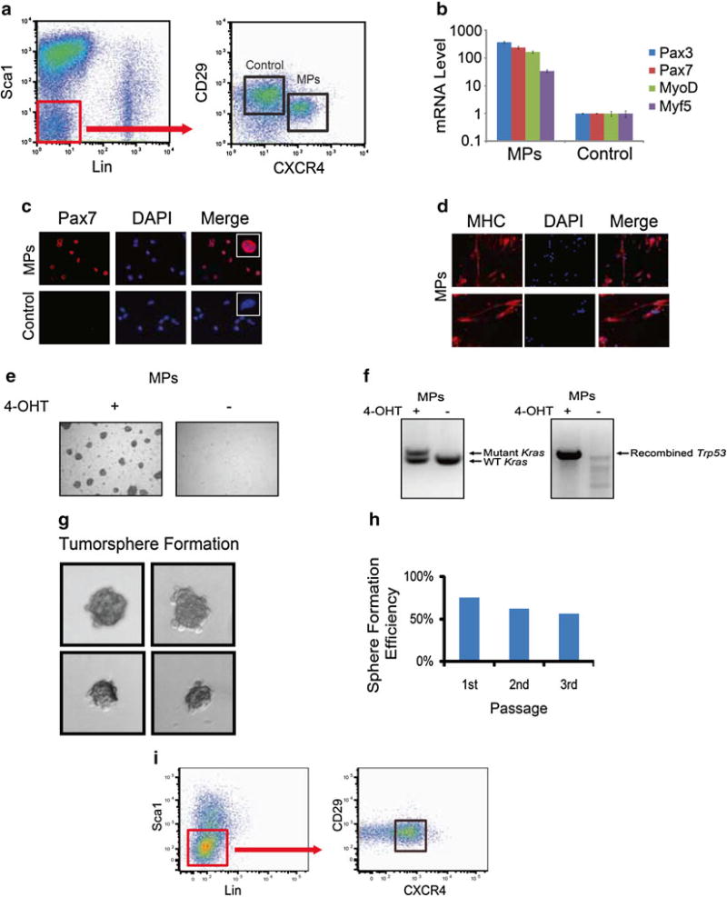
Fig. 1.
Isolation and transformation of Pax7+ myogenic progenitors in vitro. (a) Representative FACS plot of the isolation of Pax7+ myogenic progenitors (MPs) by the immunophenotype CD45−, Ter119−, Sca1−, Mac1−, CXCR4+, CD29+, in R26CE/+; LSL-KrasG12D/+; Trp53Fl/Fl mice. (b) Sorted MPs express high levels of myogenic transcripts in comparison to non-myogenic control cells. (c) Pax7 immunofluorescence demonstrates Pax7 expression in the nucleus of sorted MPs, but not control cells (scale bar = 50 µm). Inset shows magnification of a single cell. (d) Sorted MPs cultured in differentiation media express myosin heavy chain (MHC). (e) Myogenic progenitors (MPs) from R26-Cre-ERT2; LSL-KrasG12D/+; Trp53Fl/Fl mice treated with 4-OHT are more proliferative and capable of forming colonies compared to untreated MPs, indicating transformation in vitro. (f) For MPs treated with 4-OHT, genomic DNA was isolated from myogenic progenitors with or without 4-OHT treatment for 14 days. PCR demonstrates the recombination of the mutant Kras allele as well as recombination of Trp53. (g) Single transformed myoblasts were sorted into 96-well plates with proliferation medium and formed tumorspheres. Fourteen days later, about 75 % of the wells contained tumorspheres. (h) Single cells from primary tumorspheres were capable of generating new tumorspheres over three passages. (i) Transformed cells maintain satellite cell immunophenotype after multiple passages

