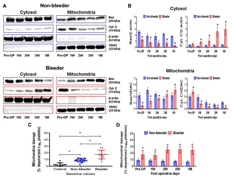
Figure 5.
Changes in Bax translocation and Cyt.C release in cytosol and mitochondrial fractions (A,B) and mitochondrial damage (C,D) in non-bleeder vs bleeder platelets at baseline and multiple time frames after CF-LVAD implantation. VDAC (for the mitochondrial fraction) and β-actin (for the cytosolic fraction) were used as the respective internal controls. The bars in the histograms represent the mean ± SD. *p<0.05 is considered significant.

