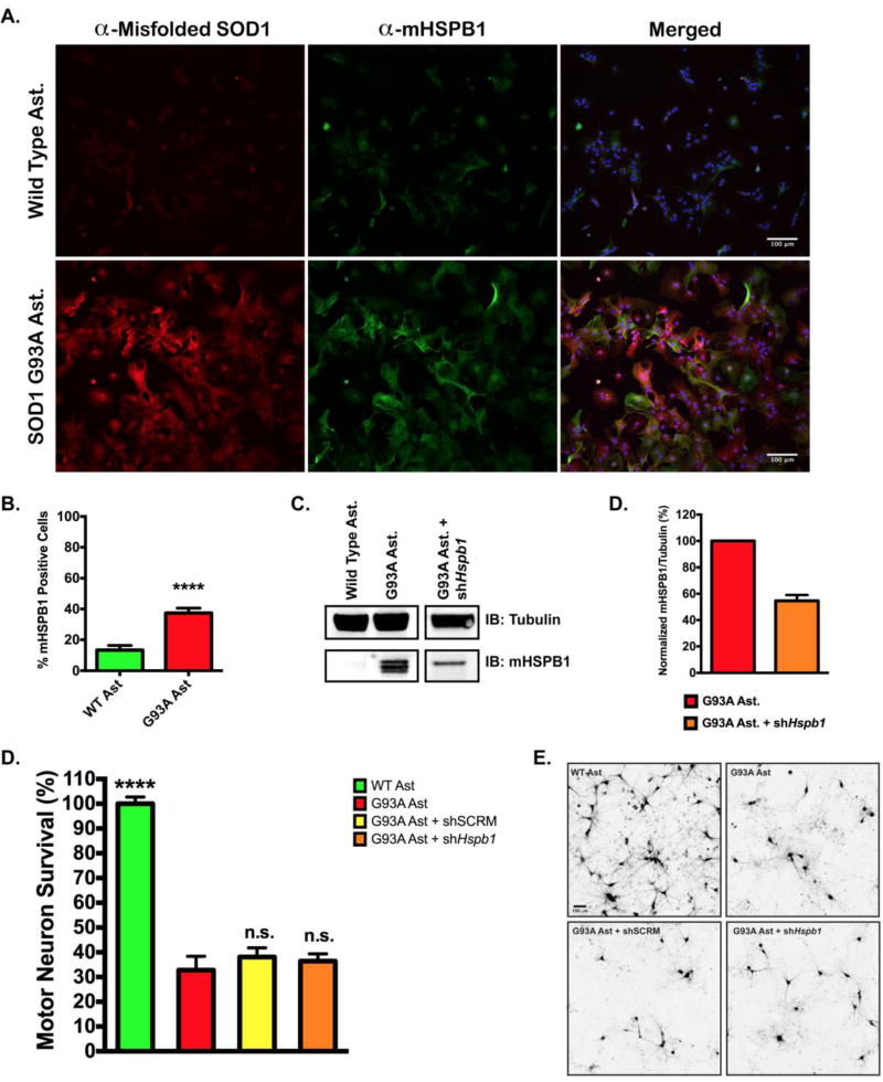
Figure 2. Knockdown of murine HSPB1 in SOD1(G93A) astrocytes has no effect on motor neuron survival.
A. Immunofluorescent staining of murine HSPB1 (mHSPB1) and misfolded SOD1 in wild type and SOD1(G93A) mouse astrocytes. Nuclei were visualized with DAPI. B. Percentage of wild type or SOD1(G93A) astrocytes stained positive for mHSPB1. Cells were counted in 5 random fields of view and expressed as the number of mHSPB1 positive cells over total cells. C. Representative immunoblot of mHSPB1 protein levels in wild type and SOD1(G93A) mouse astrocytes (left panel) and SOD1(G93A) astrocytes infected with lentiviral shRNA against Hspb1. D. Quantification of mHSPB1 immuno-staining in SOD1(G93A) astrocytes and SOD1(G93A) astrocytes transduced with shHspb1. mHSPB1 signal intensity was normalized to mHSPB1 signal intensity from SOD1(G93A) astrocyte lysates. Error bars denote s.e.m. E. MN survival at day 6 of MN co-culture assay with wild type, SOD1(G93A), SOD1(G93A) + sh Hspb1 RNA or SOD1(G93A) + shSCRM RNA astrocytes. MN survival was normalized to counts from MNs cultured with wild type astrocytes (N = 3 for all groups, each n was run in triplicate). Error bars denote s.e.m. ****P<0.0001, ns, non-significant. F. Representative images of HB9-GFP expressing MNs (shown in black) after 6 days in co-culture with wild type astrocytes, SOD1(G93A) astrocytes, and SOD1(G93A) astrocytes transduced with shSCRM or shHspb1.

