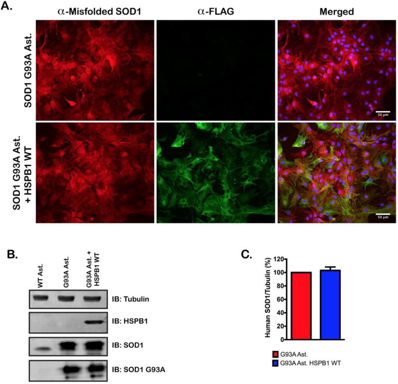
Figure 4. Overexpression of HSPB1(WT) does not reduce SOD1(G93A) expression.
A. Immunofluorescent staining of FLAG and, misfolded SOD1 in non-transduced SOD1(G93A) astrocytes and, HSPB1(WT) transduced SOD1(G93A) astrocytes. Nuclei were visualized with DAPI. B. Representative immunoblot of hHSPB1, SOD1 and misfolded SOD1 protein levels in wild type, SOD1(G93A) and SOD1(G93A) + HSPB1(WT) cell lines. HSPB1 and misfolded SOD1 were stained with antibodies specific to human isoforms of the respective proteins. SOD1 was stained using an antibody that detects both human and murine SOD1. C. Quantification of misfolded human SOD1 staining in SOD1(G93A) and SOD1(G93A) + HSPB1(WT) cell lines. Band intensities were quantified and normalized to a tubulin loading control. Data represents the average of 3 independent western blots. Error bars denote (s.e.m.) n.s., not significant.

