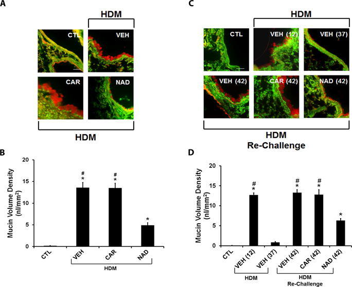
Figure 3. Effect of β-blockers on airway mucin content in the ‘prophylactic’ and ‘therapeutic’ HDM models.

Airway sections from mouse lungs were stained with periodic acid fluorescent Schiff’s stain (PAFS) for mucin (red) content in airway epithelia (green). (A and B) Mucin stained images of airway sections from the (A) ‘prophylactic’ and (B) ‘therapeutic’ models. (C) Morphometric quantification of mucin volume density in the ‘prophylactic’ model from HDM challenged mice treated with vehicle, carvedilol or nadolol in comparison to saline control mice. (D) Morphometric quantification of mucin volume density in the ‘therapeutic’ model from saline control mice compared with mice challenged with HDM and evaluated on days 12 and 37; and vehicle, carvedilol or nadolol treated mice re-challenged with HDM and evaluated on day 42. Data are mean ± SEM from 5–8 mice in each group. * represents significance at p<0.05 compared to respective saline control mice. # represents significance at p<0.05 compared to respective nadolol treated mice.
