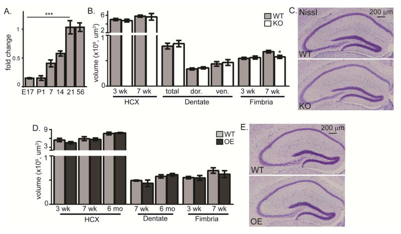
Figure 1. KL does not affect hippocampal development.
A. Brain KL qPCR fold change after normalization to 18s ribosomal subunit and adult brain KL (P56). KL was detected at embryonic (E) day 17 and postnatal (P) days 1, 7, 14, 21, and 56. B. Quantification of 3 and 7 week WT and KO hippocampus (HCX) and fimbria volume and 7 week dentate gyrus (−1.22mm to −3.88mm from bregma; dor (dorsal:−1.22mm to −2.18mm from bregma), ven (ventral: −2.20 to −3.88mm from bregma)). C. Representative 7 week WT and KO Nissl stain. Scale bar is 200μm. D. Quantification of 3 and 7 week WT and OE hippocampus (HCX) and fimbria volume and HCX and dentate volume at 7 weeks and 6 months, as in B. E. Representative 7 week WT and OE Nissl. (n=6; mean +/− S.E.M.; T-test: *p<0.05; mRNA: ANOVA, Dunnett’s post hoc analysis,***p<0.0003 relative to P56).

