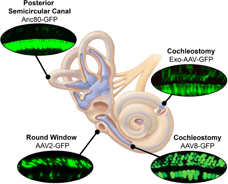
Fig. 1.
Schematic diagram illustrating several routes of delivery for injecting gene therapy vectors into the inner ear. Fluorescent images showing expression of green fluorescent protein were selected from the literature to illustrate use of several AAV vector serotypes and delivery routes from the top right, moving clockwise, they are as follows: cochleostomy, exo-AAV-GFP (György et al. 2017); cochleostomy, AAV8-GFP (Kilpatrick et al., 2011); round window membrane, AAV2-GFP (Akil et al. 2015); posterior semicircular canal, Anc80-GFP (Suzuki et al. 2017). Each fluorescence image is oriented with a single row of inner hair cells at the bottom and three rows of outer hair cells at the top.

